A visible spot or brief sensitivity does not tell you whether decay needs a filling, fluoride-based monitoring, or treatment for an injured pulp. By the end, you will know which findings dentists use to judge depth, when a filling is suitable, and which questions to ask before treatment begins.
Key takeaways
- Noncavitated white or brown spots may respond to fluoride instead of drilling.
- A visible hole, soft dentine, or progressing decay usually needs a filling.
- X-rays and pulp tests help assess decay depth and nerve involvement.
- Ask about material choice, remaining tooth structure, and alternatives before treatment.
Is the lesion still early enough for fluoride, or does it need a filling?
An intact enamel surface with a chalky white or brown spot is not automatically a cavity needing a filling. An early enamel lesion, or noncavitated lesion, has not formed a hole.
If it is smooth and hard, it may be arrested or remineralized with twice-daily brushing using 1,000–1,500 ppm fluoride toothpaste, reduced sugar frequency, professional fluoride treatment, and monitoring.
A rough, matte spot covered with plaque suggests active decay because its surface is harder to clean. A cavitated cavity is different: the defect traps plaque, and brushing alone cannot reliably keep it clean. That shifts the decision toward restoration, based on its activity, location, cleansability, and extent.
The word cavity does not reveal depth. Decay can remain limited to enamel, extend into dentin as dentin decay, or involve an exposed root surface. Root-surface caries needs separate assessment because root dentin is less mineralized and can progress faster than enamel.
Ask the dentist to check:
- Is the surface smooth and hard, or rough, matte, and plaque-retentive?
- Is there an actual opening that traps plaque?
- Could a selected noncavitated lesion between teeth be treated with resin infiltration, which slows progression without a conventional filling?
- Is the lesion on exposed root dentin, where fluoride, plaque control, and management of root exposure may be needed before deciding on restoration?
A visible dark mark alone does not prove that a filling is necessary. Surface condition and cleansability matter more.
How does a dentist determine whether decay is near the pulp?
A dentist determines proximity to the pulp by combining examination findings, imaging, and pulp testing. No single result proves how deep the decay is.
1. The dentist visually inspects the tooth after cleaning and drying it, then gently probes the surface for roughness, softness, or a true opening. They check whether plaque collects there and whether brushing, flossing, or an interdental brush can keep the area clean.
2. A bitewing X-ray examines decay between teeth and estimates whether an adjacent-surface lesion has entered dentin. A periapical X-ray is added when the dentist needs to review the entire root, the root tip, or the surrounding bone.
3. Pulp testing uses cold, heat, or electric stimulation to assess whether the nerve responds and whether that response is normal. The dentist compares the result with nearby teeth and considers how quickly the sensation stops.
4. The dentist combines these findings with lesion activity: a chalky, plaque-covered, progressing area raises different concerns from a hard, inactive lesion. An X-ray estimates lesion extent; it does not measure the cavity directly or show every softened area.
Pain is not a depth gauge. A large cavity can be painless, while a smaller defect can cause sensitivity.
Seek prompt assessment for:
- Lingering hot or cold pain, including lingering sensitivity after the stimulus is removed
- Spontaneous toothache or pain that wakes you at night
- Pain on biting
- Swelling, a gum boil, or facial swelling
These signs raise concern about pulpal or periapical disease, where a simple filling may no longer be sufficient.
What makes a filling suitable once decay has been found?
A filling is suitable when decay has created a defect that is active, cavitated, and difficult to keep clean, yet enough strong tooth remains to support a sealed restoration. The decision is not based on an X-ray finding alone; filling suitability depends on:
- Whether the lesion is cavitated and active
- Cavity depth and size
- Its location, including an exposed root surface
- The amount of remaining tooth walls
- Any crack in the tooth
- Whether the dentist can control moisture and create a durable seal
A noncavitated dentin lesion can sometimes be monitored or managed with fluoride, plaque control, sugar-frequency changes, or microinvasive care when you can attend reliable follow-up. An active cavity with a rough, plaque-retentive opening is different: brushing may not reach its walls, so leaving it unrestored becomes harder to manage predictably.
Deep decay needs a careful balance. With selective caries removal, the dentist removes infected tissue and creates a clean peripheral seal while leaving softened or affected dentin near the pulp when necessary. Removing every softened area can expose the pulp and turn a restorable tooth into one needing more extensive treatment.
A simple filling is no longer the likely answer when there is:
- Lingering or spontaneous pulpal pain
- Infection, swelling, or a gum boil
- An unrestorable crack
- Too little sound tooth structure
Those findings can lead to root canal treatment, a crown-based plan, or extraction.
What happens during the filling appointment, and which material fits the tooth?
A filling appointment starts with examination and treatment planning, including the cavity’s size, depth, location, remaining tooth structure, cracks, bite force, and ability to control moisture.
- You receive local anaesthetic when drilling could cause discomfort.
- The dentist places a rubber dam or uses cotton rolls, suction, and cheek retractors to isolate the tooth.
- Infected, softened tooth structure is removed conservatively; removing every softened area in a deep lesion can expose the pulp.
- The chosen material is placed directly, or an impression or scan begins an indirect restoration.
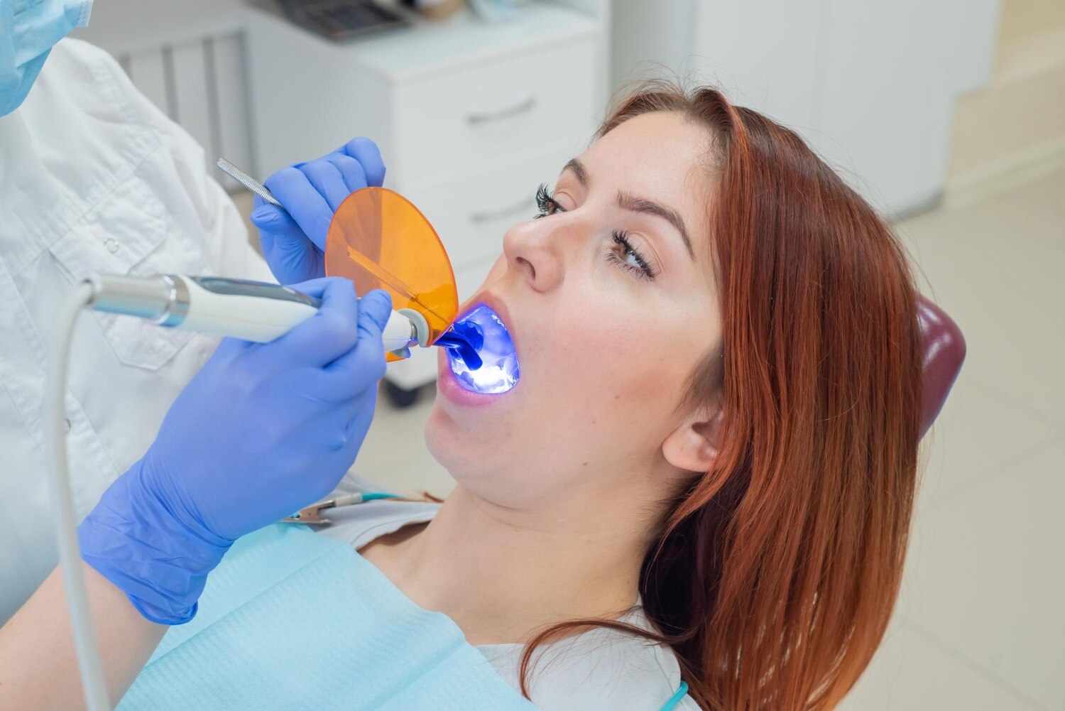
- Composite resin and some glass ionomer materials are light-cured in layers when required.
- The dentist checks your bite with articulating paper and adjusts any high spot.
- The restoration is polished, and the margins are checked.
| Option | Main advantage | Main trade-off | Common consideration |
|---|---|---|---|
| Composite resin | Tooth-coloured and adhesive | Requires careful moisture control | Useful where appearance matters and isolation is reliable |
| Glass ionomer | Releases fluoride and tolerates moisture better | Lower wear resistance in high-load areas | Suits selected low-stress sites or moisture-challenging conditions |
| Amalgam | Long record of strength in posterior teeth | Silver-coloured | Requires assessment of remaining tooth structure and current clinical indications |
| Indirect restoration | Inlays, onlays, or crowns can protect larger defects | Requires extra appointments or laboratory work | Fits a large cavity when a direct filling would leave weak walls |
Front versus back teeth, cavity size, biting force, and isolation can change the recommendation. No material is universally best: a dentist matches durability, appearance, moisture control, and remaining tooth strength to that specific tooth.
What should you ask before deciding on a filling?
Before agreeing to a filling, ask questions before a filling that establish whether treatment is necessary and durable:
- Is the surface intact or cavitated, and is the lesion active?
- Can you keep it clean with brushing, or does it trap plaque?
- Does the bitewing show enamel-only or dentin involvement?
- Did the pulp respond normally to cold, heat, or electric testing?
- How much sound wall remains, and is there a crack?
- Which material is indicated for this tooth, and why?
- What happens if the lesion is monitored instead?
A noncavitated lesion may suit fluoride, resin infiltration, or caries monitoring. A cavitated, active defect that cannot be cleaned is harder to manage without restoration. Ask for a review plan, brushing and fluoride instructions, sugar-frequency changes, and advice on checking restoration margins; a filling does not stop caries elsewhere.
Material choice depends on location, moisture control, bite force, and remaining walls:
| Option | Main trade-off | Ask when |
|---|---|---|
| Composite resin | Tooth-coloured but moisture-sensitive | The field can stay dry |
| Glass ionomer | Fluoride release and better moisture tolerance, but lower wear resistance | Moisture control is difficult or load is limited |
| Indirect inlay, onlay, or crown | More laboratory work, useful for larger weak defects | A direct filling would leave fragile walls |
Children’s primary molars need separate judgment. The Hall technique seals selected carious molars under a preformed metal crown but is unsuitable with pulpal or periapical disease. When comparing consultations, including one at Dental Junction, judge the reasoning, alternatives, and follow-up care—not the material name alone.
Related service
Tooth Filling Tooth filling is a common dental procedure used to repair teeth affected by cavities or minor damage. View service → |
Frequently asked questions
Can fluoride treat an early tooth lesion instead of a filling?
Yes, when the enamel lesion is noncavitated and has no hole, fluoride, plaque control, and dietary changes may stop or reverse progression. A cavitated lesion cannot be restored with fluoride alone.
How does a dentist determine whether decay is near the pulp?
The dentist combines visual and tactile examination with bitewing X-rays when needed. Cold testing, percussion, and symptoms such as spontaneous or lingering pain help assess pulp involvement.
What makes a filling suitable after decay is found?
The dentist considers lesion size, depth, location, moisture control, bite forces, remaining tooth structure, appearance, and your risk of future decay.
What happens during a tooth-filling appointment?
The dentist numbs the area, isolates the tooth, removes infected tooth structure, cleans the cavity, places the chosen material, shapes the restoration, and checks your bite.
What should you ask before deciding on a filling?
Ask whether the lesion is cavitated, how deep it is, whether fluoride treatment is appropriate, which materials fit the tooth, and what risks follow if treatment is delayed.